


Imaging tissue oxygenation to improve medical treatment
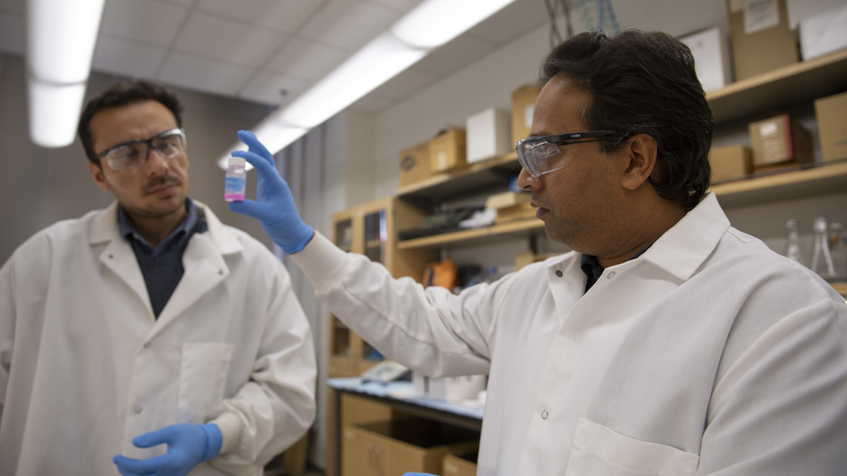
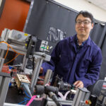
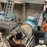
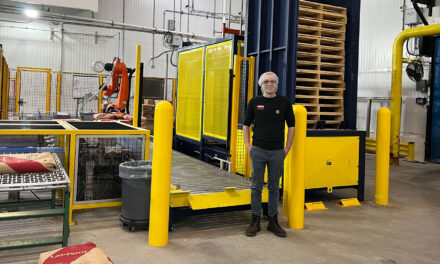
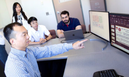
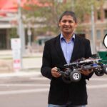
<p style="font-size:12px; margin: 0px 100px 0px 100px; text-align:center; font-style:italic">Above: Vikram Kodibagkar (right) with graduate student, Babak Moghadas examines a dual modality siloxane core nanoemulsion that is used to label implantable cells and measure the oxygenation of tissue/cells with MRI. Photographer: Erika Gronek/ASU</p>
Oxygen is at the center of everything. It’s what we breathe, and it powers our organs through the process of tissue oxygenation. Understanding this process could be the key to more effective cancer treatments.
For almost four years, Vikram Kodibagkar, an associate professor in the Ira A. Fulton Schools of Engineering at Arizona State University, has been researching a new method for measuring tissue oxygenation using advanced 3D imaging. His goal has been to further develop non-invasive techniques to measure tissue oxygenation by turning currently used qualitative techniques into quantitative data.
Kodibagkar’s research, funded by a 2014 National Science Foundation CAREER Award, is helping find ways to quantify the amount of oxygen present in tissues, or tissue oxygenation, using quantitative data. This research opens a range of applications, including improved cancer treatments.
In many cases, as a cancerous tumor grows, it rapidly outgrows its blood supply, leaving portions of the tumor with areas where the oxygen concentration is significantly lower than in healthy tissues. These oxygen-deficient, or hypoxic, areas indicate tumor cells that have been deprived of oxygen.
“Knowing about the oxygenation [of tissues] might allow us to tailor the therapy to make it better and to use other therapeutic interventions that are more appropriate,” Kodibagkar said. “There are new therapies that can target the hypoxia.”
Standard therapies used to treat tumors, like radiotherapy, are not optimally effective in hypoxic regions. However, armed with a new tissue oxygenation measuring technique to provide information about the locations of hypoxic areas, radiologists can boost the dosage of radiotherapy where tumors pose the greatest risk of metastasizing (spreading cancer cells throughout the body) or resisting therapy.
While working to improve tissue oxygenation, Kodibagkar found that the application could also be used for other therapies, including rehabilitating patients with traumatic brain injuries or those dealing with the aftereffects of stroke or heart attacks.
“We’re discovering [the research] could be useful not just in the original cancer context,” Kodibagkar said. “It’s becoming more useful as we are learning about primary brain injuries, as well as understanding oxygenation in the [healthy] brain in general.”
Kodibagkar leads the Prognostic Bioengineering (ProBE) Lab at ASU, which has a mission to develop new imaging technologies that detect changes in tissue microenvironments and training new leaders in biomedical imaging.
How the new technique works
A contrast agent is a substance used in imaging that allows researchers to see specific structures or fluids within the body. The perfusion of the contrast agent — how it passes through tissue — and its retention in hypoxic areas is the key to the technique’s success.
While the contrast agent is in the extracellular, extravascular space – outside of both the blood vessels and cells – researchers document the how much it binds to its environment.
If a contrast agent accumulates in a region of tissue, researchers can infer it has passed through “leaky,” more damaged vessels, which are commonly found in tumors or as a result of traumatic brain injury.
If the team’s newly developed contrast agent is retained for a period of three hours or more, researchers can infer the presence of hypoxia in the tissue.
“Based on the time course of the agent, or rather the concentration, now we can tie that to actual pharmacokinetic models of how molecules behave when injected into the bloodstream,” Kodibagkar said.
Using Magnetic Resonance Imaging scanners, researchers can watch for concentrated areas of a contrast agent over time to see how it moves through the vasculature (the arrangement of blood vessels in the body).
Because nearly all hospitals have access to MRI scanners, Kodibagkar’s technique could be widely accessible to clinical teams treating cancer patients and other illnesses that lead to changes in tissue oxygenation.
His research team uses pharmacokinetic modeling to detail the contrast agent’s movement, employing 3D modeling calibration phantoms to simulate different conditions in the body, such as temperature and the amount of oxygen present. The phantoms are scanned to evaluate the performance of an imaging agent and correlate its results with controlled environmental changes.
The data collected are fit to a pharmacokinetic model to extract quantitative information about oxygenation based on the imaging data. Researchers can then compare results using other techniques to validate their data.
Early in the project, the team had to redesign the calibration phantom and synthesize their own contrast agent for cost efficiency, as purchasing the contrast agent custom-made commercially would be too expensive.
“One unexpected takeaway message for the world is that we’ve developed an agent that does both perfusion and hypoxia imaging,” Kodibagkar said. “In that sense, it would be superior to a commercial contrast agent.”
Developing the new agent supports his CAREER award research goals by facilitating testing of the calibration phantoms and improving pharmacokinetic modeling using 3D imaging.
Introducing students to imaging research
In movies and television, science is everywhere. In “Jurassic Park,” for instance, advanced genetic engineering brings dinosaurs back to life. In “Back to the Future,” theoretical physics helps create time travel. But actual biomedical imaging science and engineering are rarely depicted on the screen.
“In school, you don’t really think about imaging as a career option,” Kodibagkar said. “There might be a TV show or a movie that shows it, but nobody really talks about it as a career option. From my perspective, imaging research is a very fulfilling and impactful career.”
Because many young students are unfamiliar with this relatively obscure field and less likely to pursue a career in imaging, Kodibagkar is partnering with the Fulton Undergraduate Research Initiative, known as FURI, to introduce students to career options in imaging research.
Kodibagkar has designed an eight-week Hands-on Summer Program in Imaging Technology, called HoSPIT. In the program, high school, undergraduate and master’s-level students are introduced to careers in imaging and its applications.
A group of high school students completed the first HoSPIT program in summer 2014. Since then, 16 high school and undergraduate students have also completed it.
“The work I did with Dr. Kodibagkar has provided me with the strong foundation not only in the field of imaging, but also as a scientist for conducting good quality research in the future,” said Shubhangi Agarwal, who recently completed a biomedical engineering doctoral degree program.
Agarwal is now a postdoctoral researcher at the University of California at San Francisco. She continues to apply the MRI techniques she learned at ASU to study cancer metabolomics.
Kodibagkar’s research “was the perfect opportunity for me to venture into the field of cancer treatment and imaging,” Agarwal said. “MR cancer imaging has the potential to provide complex information about cancer’s microenvironment without the need for extensive, invasive methods.”
Kodibagkar has also partnered with a colleague in the School of Biological and Health Systems Engineering, Assistant Professor Barbara Smith, on a global summer internship program she leads to recruit undergraduate students from India. Before the project concludes in the spring, Kodibagkar will mentor another doctoral student, Babak Moghadas, through completing key research goals of the project.
“The biggest impact of the NSF CAREER Award has been on the students who received training in cutting-edge imaging techniques under its auspices and who represent the next generation of leaders in imaging,” Kodibagkar said.

