Bonding experience: Repairing wounds with gold, silk and lasers
New nanomaterial-welding method raising hope of a safer way to heal damaged body tissues and surgical incisions
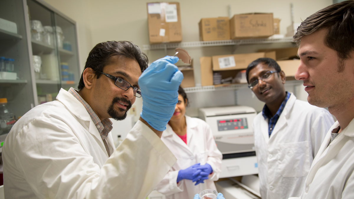
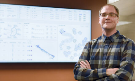
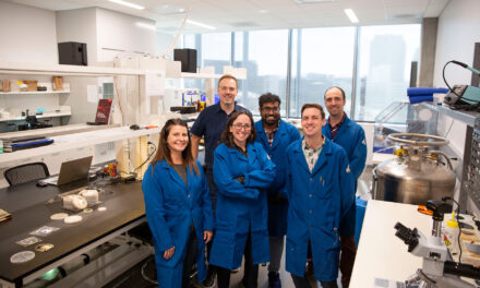
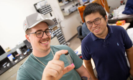
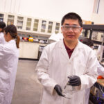
Above: Professor Kaushal Rege directs student research assistants in his Molecular and Nanoscale Bioengineering Lab at Arizona State University. Rege is working with Professor Jeff Yarger and his research group in ASU’s School of Molecular Sciences, and Jacquelyn Kilbourne, assistant director of ASU’s Department of Animal Care and Technologies, to develop a safer, more effective technique for sealing and healing body tissues. Photographer: Nora Skrodenis/ASU
A dermal regenerator was the medical tool used to repair spaceship crew members’ wounds on the science fiction TV show “Star Trek.”
Another kind of tissue-repair technique emerging from Arizona State University Professor Kaushal Rege’s research has been compared to the fictional device that healed damaged flesh by simply being passed over a wounded body part.
The comparison is a bit of a stretch, says Rege, who teaches in the School for Engineering of Matter, Transport and Energy, one of the six Ira A. Fulton Schools of Engineering at ASU. But Rege and his research collaborators are excited about their new tissue-bonding method utilizing the restorative capabilities of nanomaterials.
Over the past five years, the team in Rege’s Molecular and Nanoscale Bioengineering Lab has been experimenting with this laser-activated process that promises to be effective as a reinforcement for conventional stitches used to seal wounds and surgical incisions, and in some cases provide a safer, more resilient alternative.
Interlacing molecules to create a ‘bioactive’ sealant
The biggest advance in the project, which has funding from the Mayo Clinic and the National Institute of Biomedical Imaging and Bioengineering, is the discovery of a powerful combination of two long-treasured materials that make this nanomaterial-welding method an effective tissue sealant — silk and gold.
Using silk produced by silkworms and tiny gold nanorods (roughly 10,000 times smaller in diameter than a single human hair), researchers zap the nanorods with laser light, causing electrons in the gold to oscillate.
“Those oscillations produce a rise in temperature caused by the release of energy from the gold nanorods as they convert the laser light into heat,” Rege says.
That heat can then produce changes in the material properties of the silk and collagen, the main structural protein found in skin and other connective tissues.
When the heat dissipates, the tissue molecules and the silk molecules “interdigitize,” Rege says. “They essentially interlace,” creating “bioactive sealing.”
So, in other words, the heated laser light essentially welds together silk molecules and tissue molecules to close an open wound or incision.
Testing the capabilities of tissue-bonding materials
That progress has set the stage for answering crucial questions: Can the biomechanical properties involved in the laser, gold and silk bonding technique provide the necessary strength to hold all the various kinds of body tissues together, and how long can the bonding endure?
Rege points out that the method is working on skin, but he and his team are still exploring its use when applied to the tissues of internal organs. Other formulations or mixtures of different materials might be necessary to expand the technique’s capabilities to sealing wounds or incisions in and around internal organs, he says.
“The materials have to withstand the kinds of the biomechanical stresses that occur in delicate places in the body, such as in colorectal tissue,” Rege says.
The bonding materials also have to be tested to prove they can be both nontoxic and biodegradable in all applications.
“The main thing is that the sealing has enough mechanical strength so that leaks and separations and infections don’t happen and the body’s natural healing processes have time to take over,” he says, “and can we deliver bioactive molecules to the site to facilitate that healing?”
Next step: Expanding the sealing method’s biomedical applications
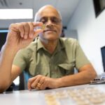
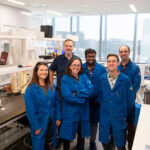
Rege’s key collaborators are Professor Jeff Yarger and his research group in ASU’s School of Molecular Sciences, whose expertise is in silk and related biopolymers, and Jacquelyn Kilbourne, assistant director of the Department of Animal Care and Technologies at ASU. Yarger and Kilbourne’s contributions are important to paving the way for safe clinical testing of new tissue-repair materials and methods, Rege says.
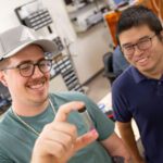
Chemical engineering doctoral student Russell Urie and biological design doctoral student Deepanjan Ghosh are Rege’s research assistants on the project. Chengcheng Guo, a former graduate student in Yarger’s silk research lab, has also aided Rege with the research.
Rege’s lab team is also working to determine the effectiveness of the laser-sealing approach in fighting and preventing infections at the sites of surgical incisions. This work could prove to be important in fighting off hospital-acquired infections such as methicillin-resistant Staphylococcus aureus, commonly called MRSA. The bacterium causes infections in different parts of the body and can lead to serious complications, especially in immune-compromised individuals.
While the research endeavor has progressed beyond basic development and testing, the team must now undertake more challenging clinical trials and make technological refinements. Once those steps are completed, Rege says he’s confident the technique will prove valuable in multiple biomedical applications, including nerve and cardiovascular surgery and repair.


