Applying medical imaging expertise to battles against kidney disease, nervous system disorder
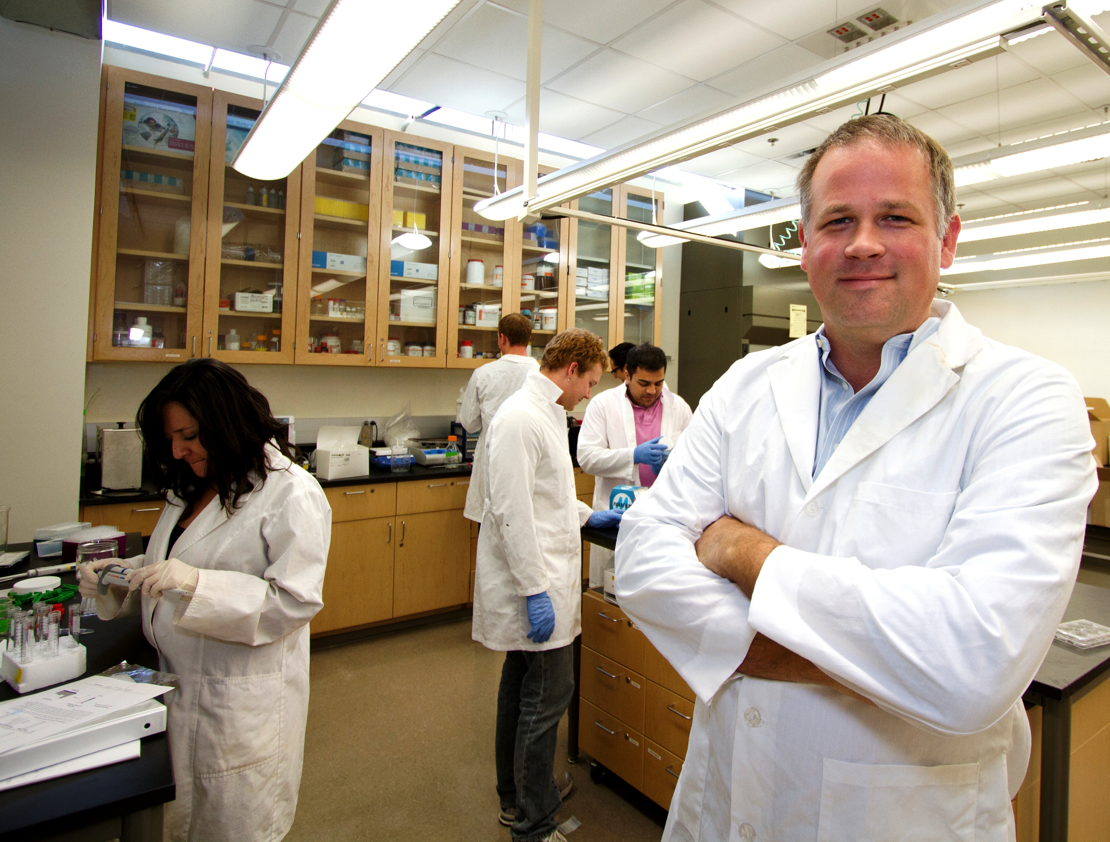
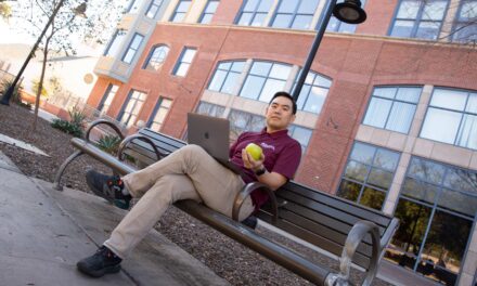
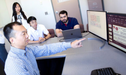
Biomedical engineer and physicist Kevin Bennett works with ASU engineering students and colleagues at the Barrow Neurological Institute and Mayo Clinic Scottsdale on research to fight kidney disease and neurofibromatosis.
Posted: February 21, 2012
Promising efforts to improve detection of early-stage kidney disease and treat children with neurofibromatosis have earned grants for Arizona State University research projects from the American Heart Association (AHA) and the National Institutes of Health (NIH).
Kevin Bennett, a biomedical engineer and physicist at ASU, is playing a leading role in both projects. Bennett is an assistant professor in the School of Biological and Health Systems Engineering, one of ASU’s Ira A. Fulton Schools of Engineering. He also is the undergraduate program chair for the school’s Harrington Bioengineering Program.
His work focuses on medical imaging, specifically the development and application of magnetic resonance imaging (MRI). He has conducted post-doctoral research in the area for the National Institutes of Health.
“I love developing new ways to see things in the body that we weren’t able to see before,” he says.
For the past five years, Bennett has been using MRI to examine kidney structure and function and to detect early stages of kidney diseases.
He and his research team use “magnetic nanoparticles and super high-field MRI to make very precise measurements of kidney structure and function,” says Bennett, who is also an adjunct assistant professor of radiology at Mayo Clinic Scottsdale.
A kidney’s susceptibility to disease can be determined by examining MRI images and determining the amount of nanoparticles that collect in a kidney’s filtering nephrons. Nephrons regulate the levels of water and soluble substances in the blood.
MRI can be used to examine the functionality of nephrons in living organisms to assess risk of kidney disease, as well as “to help measure how well a donor kidney is going to function once it’s transplanted,” Bennett says.
The AHA recently awarded $140,000 to help continue the project. Bennett’s grant proposal was among the 13 percent of requests to be approved for funding.
The grant recognizes the value of his team’s work, he says, because the AHA typically selects projects it considers promising to make breakthroughs and have a significant impact on human health.
Bennett is working on the project with John Bertram, a professor and head of the Department of Anatomy and Developmental Biology at Monash University in Australia, and Teresa Wu, an associate professor of industrial engineering and director of the Collaborative Decisions Lab at ASU, and an associate professor of radiology at the Mayo Clinic College of Medicine.
Also on the team is ASU biomedical engineering doctoral student Scott Beeman and industrial engineering doctoral student Min Zhang.
Bennett is collaborating with Vinodh Narayanan, a pediatric neurologist and researcher with the Barrow Neurological Institute (BNI) in Phoenix and adjunct faculty member at ASU, to find a drug that can reverse the effects of cognitive deficit symptoms in children with neurofibromatosis.
Neurofibromatosis is an incurable genetic disorder of the nervous system whose symptoms range from tumors to bone disorders. Narayanan, the lead researcher for the project, works with patients who have cognitive deficits caused by a specific gene, which leads to neurofibromatosis.
It’s been proposed that the cognitive deficits in people with the condition are caused by “a certain kind of molecular transport in cells that is being blocked,” Bennett says.
Neuron cells have an axon – a long nerve fiber that transports electrical impulses. Narayanan believes that the axonal transport is what is blocked by neurofibromatosis. He is targeting these axons in his experiments.
As a co-investigator, Bennett is helping by using MRI to view and record the effects of certain drugs targeted to increase the transport rates of cells. He’s introducing manganese ions to cell transport because the ions can brighten MRI images
Manganese ions behave like calcium in cells and follow the same transport paths. So when paired with MRI, the manganese allows researchers to track the rate at which axons are transporting matter through cells.
“We just squirt a little manganese into the nose and we monitor how fast manganese is moved from the nose into the olfactory bulb in the brain,” Bennett says.
The process is then used in combination with certain drugs to test their ability to increase the rate of transport.
Research is being performed at both ASU and the BNI, focusing mostly in Narayanan’s lab and at the BNI-ASU preclinical imaging center.
The project was making advances significant enough to attract support from the NIH. Narayanan’s team has been awarded $275,000 to continue the work. Only about 10 percent of applicants for such funding were selected to receive NIH grants.
The grant comes from R21 funding, which is reserved for high-risk, high-impact projects. Applicants compete with accomplished researchers across the country for the R21 grants, Bennett says.
“I think our success [at winning a grant] can be attributed to Dr. Narayanan’s brilliance and our productive collaboration,” he says.
The Army Research Office initially funded the project for two years with a grant of $120,000.
Bennett says it’s especially rewarding when his team can collaborate on research pursuing solutions to critical biomedical challenges.
“We get to see our work applied to important fundamental research and to clinical problems,” he says.
Written by Natalie Pierce and Joe Kullman


