


Bioimaging funding stimulates harmonized research
Fulton Schools faculty members receive support from Chan Zuckerberg Initiative for multidisciplinary research
A portmanteau of science and dialog, Scialog supports research and multidisciplinary collaboration allowing researchers to address scientific challenges of global significance. For three Arizona State University researchers, selection as grantees for Scialog: Advancing BioImaging will give them a unique opportunity to explore the next generation of imaging technologies in a three-year initiative designed to spark creativity and generate ideas for novel research projects.
Benjamin Bartelle, assistant biomedical engineering professor, and Barbara Smith, associate biomedical engineering professor, in the Ira A. Fulton Schools of Engineering at ASU, and Douglas Shepherd, an assistant physics professor in the College of Liberal Arts and Sciences, were each selected to participate on one of 10 research teams bringing together physicists, biologists, bioengineers and medical imaging specialists to develop solutions in advanced bioimaging.
Each researcher will receive $50,000 in funding to support their team’s project. The Research Corporation for Science Advancement, Chan Zuckerberg Initiative, or CZI, and the Frederick Gardner Cottrell Foundation are funding a combined total of $1.15 million to support 23 awards across 10 research teams in this inaugural year.
“Imaging technologies play a critical role in CZI’s mission to support the science and technology that will make it possible to cure, prevent or manage all disease by the end of the century,” says Stephani Otte, science program officer for imaging at CZI. “We hope these teams of early-career researchers will advance the imaging field’s ability to observe and analyze biological processes and help build a much deeper mechanistic understanding of biological systems, identify potential points of intervention in disease and inform directive treatments.”
Converging to decode data
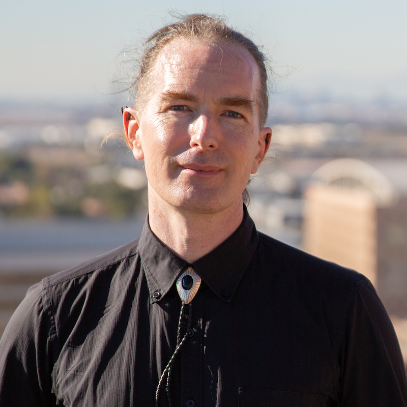
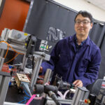
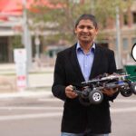
Benjamin Bartelle
The first virtual meeting of the Scialog: Advanced BioImaging series occurred in May and it’s where Bartelle and collaborators, Lu Wei of the California Institute of Technology and Ulugbek Kamilov of Washington University in St. Louis, began discussions for their project, “Enabling Noninvasive Lipid Profiling with Intermodal Deep Learning.”
Lipids are a part of every cell and embedded in every tissue in the body. Lipid biomarkers can be found in everything from blood samples to tumors, but in most cases, it is unknown if they are the cause or the product of a disease.
The team will use MR Spectroscopy and Raman imaging, techniques that generate images with both spectral and spatial information, to detect lipids. They will use the Raman imaging, which is highly sensitive, to decode MR Imaging, which can be used clinically, on the same field of view using an artificial intelligence algorithm called Deep Learning.”
Bartelle says that the goal of his lab is to create new tools to resolve and manipulate neuroimmune signaling.
“Lipid signaling is one well-known form of inflammatory signaling that we can currently detect only by drawing blood and measuring from plasma,” says Bartelle. “I don’t just say ‘inflammatory signaling’ because our best understanding of that right now is that there are dozens of different kinds of inflammation that can’t be treated the same way.”
Circulating lipids like triglycerides can indicate inflammation or heart disease; but if diseased tissue is on the other side of the blood-brain barrier, it may be difficult to detect. A patient could also have some focal inflammatory signaling that a drop of blood would not locate.
This is where MRI comes into the picture.
“We can see almost everywhere in a human patient and sometimes you can even pick up inflammation by seeing edema,” says Bartelle. “That’s a dead giveaway for something like a stroke, but that doesn’t give you any specifics on what is happening. You need something molecularly specific. It turns out MRI might be good for that, too.”
MRI is based on chemistry method nuclear magnetic resonance, which allows for information about atomic nuclei to be read out as a spectrum of molecular information. Combining the imaging of MRI with the molecular information from nuclear magnetic resonance is called magnetic resonance spectroscopic imaging, or MRSI.
“I was talking with Lu Wei about how her method, Raman spectroscopy, is actually really great at identifying all kinds of individual molecules from tissue, as long as you have a slice in a dish in front of your Raman microscope,” says Bartelle. “We wondered if there was a way to use Raman data to ‘decode’ MRSI data. Dr. Kamilov has a lot of experience building such decoders, so we approached him about working together.”
The three researchers know enough about each other’s fields to harmonize their approaches while lending expertise in their respective specialties. Wei and Bartelle will collect MRSI and Raman data and translate the information to assist Kamilov in the decoding process.
Bartelle says that if they achieve their goal, they will have a computational tool that can take MRSI data and translate it into individual chemical species using an algorithm trained on Raman data.
“The end result would be something you could use on a patient to diagnose what kind of inflammation is going on, where it is and how best to treat it,” says Bartelle. “Think of it as a machine learning tool that can read things from MRSI that no human could. This could become a critical tool for determining a course of treatment for any brain disorder from migraine to stroke to neurodegeneration. We’ve never been able to see these things before so it’s hard to say just what will be the ultimate application.”
The innovative thinking and fusion of ideas and expertise are why Scialog convenes scientists to explore cutting-edge projects.
Two imaging systems, one novel solution
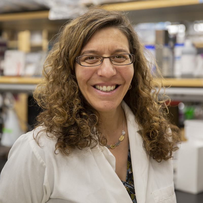
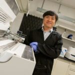
Barbara Smith
Barbara Smith’s Scialog: Advancing BioImaging award is for a project named “Microendoscopy-Guided Diagnosis and Treatment of Early-Stage Ovarian Cancer”, It is a collaboration with Bryan Spring of Northeastern University, and will draw on her research, which uses microendoscopy to diagnose and treat early-stage cancer.
Smith and Spring are currently developing independent microendoscopy systems. Their project will enable the integration of two imaging techniques to investigate early-stage ovarian cancer as it develops in the fallopian tube.
“This vision to develop practical screening methods aims to save lives and reduce health care costs by mitigating the occurrence of advanced-stage disease,” says Smith. “Through this work, we aim to develop a clinically relevant imaging tool capable of scanning the entire fallopian tube to enable routine screenings for early-stage ovarian cancer detection.”
The project could result in an innovative approach to screen a large volume area at a high resolution in the fallopian tube.
“This enables, for the first time, synthesis of comprehensive, high-resolution volumetric renders of the fallopian tube to precisely locate neoplasms for immediate image-guided ablation, biopsy collection and follow up surveillance,” says Smith. “It would be transformative for catching and treating premalignant lesions.”
The team’s proposed advance in bioimaging is driven by a critical need and represents a bold step forward that will enable clinical diagnostics for early-stage ovarian cancer. “This collaboration formed by the Scialog grant will join labs that are distinctly suited to work together towards a critical scientific advance that has not otherwise been achieved,” says Smith.
New and better tools will always be a need in health care systems, allowing for better diagnosis and information on how to care for patients. The funding of these projects has the potential to fill gaps in modern medicine.
“With more awards than any Scialog to date, this initiative is off to a great start,” says Daniel Linzer, president and CEO of RCSA. “We’re grateful for funding partnerships that enable us to seed even more projects with the potential to transform an area of science.”


